Anion Gap Normal Range: What High and Low Results Mean (2026)
Anion gap normal range is 8 to 12 mEq/L. Albumin correction, MUDPILES causes of high anion gap, delta ratio formula, and normal gap acidosis explained.
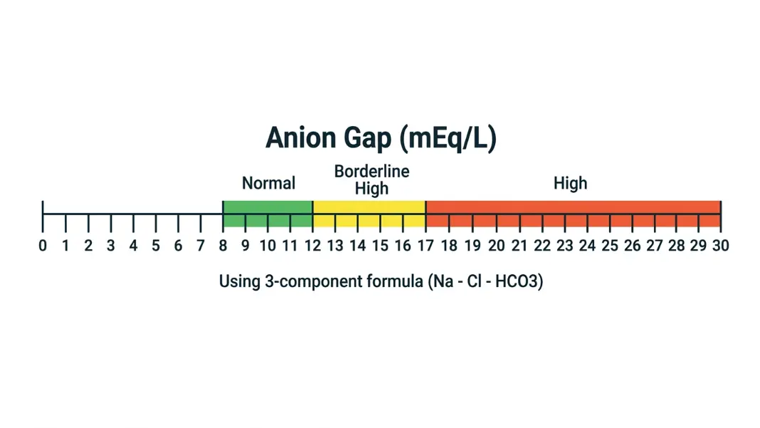
Your metabolic panel comes back with an anion gap of 18 mEq/L. The lab flags it high. Normal, according to the reference range printed on the report, is 8 to 12. But if the patient's albumin is 2.1 g/dL instead of 4.0, that number changes the interpretation before you even begin looking at causes. An uncorrected anion gap in a hypoalbuminemic patient hides a real metabolic acidosis just as easily as it creates a false high.
The Anion Gap Calculator runs the albumin-corrected value automatically from your sodium, chloride, bicarbonate, and albumin inputs. This guide explains how the formula works, what the normal range actually means, which conditions elevate the gap, and when a normal gap does not rule out acidosis.
The Anion Gap Formula: What the Number Actually Represents#
The anion gap is the difference between measured cations and measured anions in the blood. The body maintains electrical neutrality, so total positive charge in plasma always equals total negative charge. But not every ion is measured on a standard metabolic panel. The "gap" is the space filled by unmeasured anions, primarily albumin, phosphate, sulfate, and organic acids.
Anion Gap = Sodium - (Chloride + Bicarbonate)
Worked example:
- Sodium: 140 mEq/L
- Chloride: 102 mEq/L
- Bicarbonate: 24 mEq/L
- Anion Gap = 140 - (102 + 24) = 140 - 126 = 14 mEq/L
Some labs use a 4-component formula that adds potassium to sodium:
Anion Gap = (Sodium + Potassium) - (Chloride + Bicarbonate)
The 4-component version produces a result about 4 mEq/L higher than the 3-component formula. Your lab's reference range is calibrated to whichever formula they use. Comparing your result to a reference range from a different formula is one of the most common reasons patients misread their own panels.
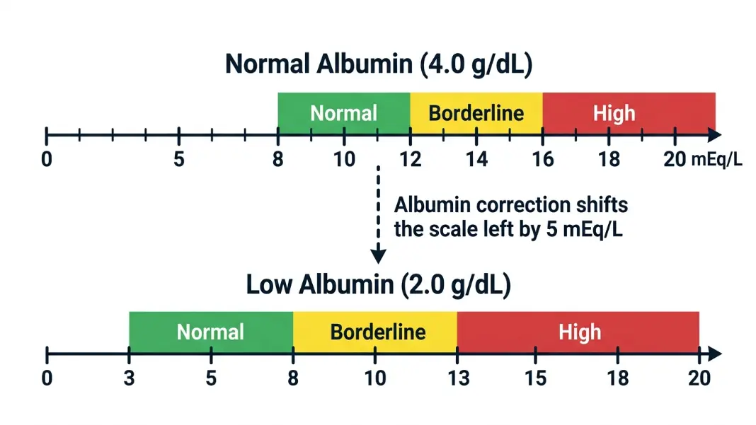
The albumin correction is not optional in hypoalbuminemic patients. Albumin is the largest contributor to unmeasured anions at baseline. When albumin falls, the gap falls with it. For every 1 g/dL that albumin drops below 4.0 g/dL, the anion gap decreases by approximately 2.5 mEq/L.
Corrected Anion Gap = Measured Anion Gap + 2.5 × (4.0 - Albumin [g/dL])
Worked example with low albumin:
- Measured anion gap: 14 mEq/L
- Albumin: 2.0 g/dL
- Correction: 2.5 × (4.0 - 2.0) = 2.5 × 2.0 = 5.0
- Corrected anion gap: 14 + 5 = 19 mEq/L
A gap that looks normal at 14 is elevated once corrected. This pattern, a falsely normal gap masking a real high-gap acidosis, appears most often in ICU patients, cirrhosis, nephrotic syndrome, and malnutrition. The Corrected Calcium Formula applies the same albumin-correction logic to total serum calcium for exactly this reason: multiple lab values only mean what they appear to mean when protein levels are normal.
Normal Anion Gap Range: Why Labs Print Different Reference Values#
The normal range for the 3-component anion gap formula is 8 to 12 mEq/L in most clinical references. Some institutions use 7 to 13 mEq/L, and labs using the 4-component formula shift the range up to approximately 12 to 20 mEq/L.
The practical implication: read your result against the reference range on your specific lab report, not a value from a textbook calibrated to a different formula.
What the normal range represents biologically: in a healthy person with normal albumin (4.0 g/dL), normal phosphate, and no organic acid accumulation, the unmeasured anion pool is stable and predictable. An anion gap of 10 reflects a normal distribution of albumin and phosphate, nothing else.
The following table shows how the expected baseline gap shifts downward as albumin falls, while the corrected normal ceiling stays the same. Skipping the correction in hypoalbuminemic patients consistently produces false negatives, not false positives.
| Albumin (g/dL) | Expected Baseline Gap | Corrected Normal Ceiling |
|---|---|---|
| 4.0 (normal) | 8 to 12 mEq/L | 12 mEq/L |
| 3.0 | 5.5 to 9.5 mEq/L | 12 mEq/L corrected |
| 2.0 | 3 to 7 mEq/L | 12 mEq/L corrected |
| 1.0 | 0.5 to 4.5 mEq/L | 12 mEq/L corrected |
A measured gap of 8 in a patient with albumin of 1.5 is not normal. The corrected value is likely above 15. The table above makes that pattern visible before you calculate.
High Anion Gap Causes: MUDPILES and What Each Category Looks Like#
A high anion gap, corrected value above 12 mEq/L, means unmeasured anions have accumulated above the expected baseline. In the presence of metabolic acidosis, a high corrected gap points toward an organic acid accumulating faster than the kidneys can clear it.
MUDPILES is the standard mnemonic for high anion gap metabolic acidosis causes:
M - Methanol Methanol poisoning occurs from ingestion of impure alcohol, industrial solvents, or bootleg spirits. It metabolizes to formic acid and formaldehyde. The gap elevation can exceed 30 mEq/L. An elevated osmolar gap in early ingestion (before full metabolism) is an additional clue.
U - Uremia End-stage kidney disease causes retention of sulfate, phosphate, and organic acid metabolites. Gap elevation is usually moderate at 16 to 24 mEq/L. Chronic uremia rarely produces the severe gaps seen in acute toxic ingestions.
D - Diabetic Ketoacidosis (DKA) DKA is the most common cause of high anion gap acidosis in emergency and inpatient settings. Ketone bodies (beta-hydroxybutyrate and acetoacetate) are the accumulating unmeasured anions. Gaps in DKA typically range from 16 to 32 mEq/L depending on severity. As ketones are cleared during treatment, watching the gap close toward normal is a useful secondary progress marker alongside glucose and bicarbonate.
P - Propylene Glycol Propylene glycol is a solvent in several IV medications, including lorazepam, diazepam, and phenobarbital infusions. High cumulative doses common in ICU sedation cause lactic acidosis and an elevated gap. This cause gets missed because the source is a prescribed medication rather than an ingested toxin.
I - Isoniazid / Iron Isoniazid overdose causes seizures and lactic acidosis. Iron toxicity causes oxidative damage and lactic acidosis through a separate mechanism. Both are less common than other MUDPILES causes but clinically significant when the history fits.
L - Lactic Acidosis Type A lactic acidosis arises from tissue hypoperfusion: septic shock, cardiogenic shock, respiratory failure. Type B occurs without overt hypoperfusion: metformin accumulation in renal failure, liver disease impairing lactate clearance, mitochondrial toxicity from certain antiretrovirals. Lactic acidosis is the most common cause of a markedly elevated gap (above 30 mEq/L) in the ICU.
E - Ethylene Glycol Ethylene glycol (antifreeze) metabolizes to oxalic acid and glycolic acid. Like methanol, early ingestion shows an elevated osmolar gap before the full acid load forms. Calcium oxalate crystals in urine are a helpful diagnostic finding when present.
S - Salicylates Aspirin overdose produces a mixed picture: respiratory alkalosis early from direct respiratory center stimulation, followed by high anion gap metabolic acidosis. A mixed pattern of respiratory alkalosis and metabolic acidosis should raise salicylate toxicity until ruled out.
In clinical practice outside the ICU, DKA, lactic acidosis, and uremia account for the large majority of cases. The MUDPILES mnemonic ensures you do not miss the rarer but reversible toxic causes, particularly methanol and ethylene glycol, which have specific antidotes and respond poorly to supportive care alone.
Normal Anion Gap Metabolic Acidosis: When the Gap Stays in Range#
A metabolic acidosis with a normal anion gap means the acidosis comes from bicarbonate loss or chloride retention rather than organic acid accumulation. The body maintains electrical neutrality by retaining chloride when bicarbonate falls, which is why this is also called hyperchloremic metabolic acidosis.
Gastrointestinal bicarbonate loss:
- Diarrhea: the most common cause overall. The small intestine secretes bicarbonate-rich fluid. High-volume diarrhea causes significant bicarbonate loss and is the first thing to consider in any outpatient presenting with a normal gap acidosis.
- Ileostomy or pancreatic fistulas produce the same pattern through continuous low-level bicarbonate loss.
- Cholestyramine and similar anion exchange resins bind bicarbonate in the gut.
Renal causes:
- Renal tubular acidosis (RTA): the tubule fails to either reabsorb bicarbonate (type 2) or secrete acid appropriately (type 1 and type 4). Each type has different associated findings and causes.
- Carbonic anhydrase inhibitors (acetazolamide) block bicarbonate reabsorption directly and are an iatrogenic cause in patients on glaucoma treatment.
- Early renal failure: before uremic anions accumulate enough to elevate the gap.
Iatrogenic: Large-volume normal saline infusion introduces a high chloride load relative to sodium, diluting bicarbonate and raising chloride. Saline-induced hyperchloremic acidosis is common after aggressive resuscitation and is often overlooked because the clinical team is focused on the primary problem that required the resuscitation.
The Corrected vs Ionized Calcium guide covers how acid-base disturbances independently affect calcium binding to albumin. A patient with normal gap metabolic acidosis and concurrent hypocalcemia symptoms may have both problems driven by the same underlying process.
The Delta Ratio: Using Both Gap Values Together#
When a patient has a high anion gap metabolic acidosis, the delta ratio tells you whether a second, overlapping process is present that you might otherwise miss.
The delta ratio compares how much the anion gap has risen above normal to how much bicarbonate has fallen below normal.
Delta Ratio = (Measured Anion Gap - 12) / (24 - Measured Bicarbonate)
Worked example:
- Measured anion gap: 22 mEq/L
- Measured bicarbonate: 16 mEq/L
- Delta ratio = (22 - 12) / (24 - 16) = 10 / 8 = 1.25
The result sits in the expected range for pure high anion gap acidosis. Here is how to interpret each zone:
| Delta Ratio | Interpretation |
|---|---|
| Below 0.4 | Normal anion gap acidosis is the primary process |
| 0.4 to 1.0 | Mixed: high gap and normal gap acidosis coexist |
| 1.0 to 2.0 | Pure high anion gap metabolic acidosis |
| Above 2.0 | High gap acidosis with concurrent metabolic alkalosis |
A delta ratio between 1 and 2 is the expected result of pure DKA or pure lactic acidosis. A ratio below 1 means the bicarbonate has fallen more than the gap has risen, which usually means a normal gap process (such as diarrhea) is running alongside the high gap acidosis.
A ratio above 2 means the bicarbonate is higher than expected for the degree of gap elevation. This happens when a metabolic alkalosis (from vomiting, diuresis, or contraction) is partially offsetting the bicarbonate drop from the acidosis. The net bicarbonate may look less alarming than the acidosis actually is.
The delta ratio functions as a red flag, not a definitive diagnosis. A number outside the 1 to 2 range is a prompt to look for a second process. What that process is requires the clinical context.
In patients with DKA specifically, both the anion gap and glucose-derived metrics are active tracking tools during treatment. The A1C Calculator provides the glucose management context, while the anion gap tracks the resolution of the ketoacidosis itself. Watching both move toward normal independently confirms that treatment is addressing both components.
The normal anion gap is 8 to 12 mEq/L using the 3-component formula: sodium minus chloride minus bicarbonate. Labs using the 4-component formula that adds potassium report a higher reference range of approximately 12 to 20 mEq/L. Albumin also shifts the expected value: every 1 g/dL drop in albumin below 4.0 lowers the gap by 2.5 mEq/L. Always compare your result to the reference range on your specific lab report, as different formulas and albumin levels change what "normal" looks like.
A high corrected anion gap (above 12 mEq/L) means unmeasured anions have accumulated in the blood above the expected baseline. In the setting of metabolic acidosis, it points toward one of the MUDPILES causes: methanol, uremia, diabetic ketoacidosis, propylene glycol, isoniazid or iron toxicity, lactic acidosis, ethylene glycol, or salicylates. In clinical practice, DKA, lactic acidosis, and uremia account for most cases. A gap above 12 without acidosis should trigger albumin correction before assuming pathology.
Anion gap (mEq/L) = Sodium minus (Chloride plus Bicarbonate). All three values come from a standard metabolic panel. If albumin is below 4.0 g/dL, apply the correction: add 2.5 times (4.0 minus albumin) to the measured gap. This corrected value is what gets compared to the normal range. Most labs report only the uncorrected gap, so the albumin correction is a manual step whenever albumin is abnormal.
Normal anion gap metabolic acidosis results from bicarbonate loss or chloride retention rather than organic acid accumulation. Diarrhea is the most common cause: the small intestine loses bicarbonate-rich fluid. Renal tubular acidosis causes the same pattern through a tubular defect in acid secretion or bicarbonate reabsorption. Large-volume normal saline infusion causes saline-induced hyperchloremic acidosis. In all these cases, chloride rises as bicarbonate falls, keeping the anion gap in the normal range.
The delta ratio is (measured anion gap minus 12) divided by (24 minus measured bicarbonate). It compares how much the gap rose above normal to how much bicarbonate fell below normal. A result between 1 and 2 indicates pure high anion gap acidosis. Below 1 suggests a coexisting normal anion gap process is also lowering bicarbonate. Above 2 suggests a concurrent metabolic alkalosis is partially offsetting the bicarbonate drop. The delta ratio flags mixed acid-base disorders that the gap and bicarbonate values alone would miss.
Yes. Albumin is the largest contributor to unmeasured anions at normal levels. When albumin falls below 4.0 g/dL, the measured anion gap falls by approximately 2.5 mEq/L for every 1 g/dL decrease. A patient with albumin of 2.0 g/dL has an expected gap 5 mEq/L lower than normal. A measured gap of 10 in that patient corrects to 15, which is elevated. Skipping the albumin correction in hypoalbuminemic patients is the most common way a real high-gap acidosis gets missed on a routine metabolic panel.
Written by
Hassaan Rasheed
Web Developer & Content Researcher
Hassaan builds calculators and writes source-linked guides across the site's subject areas. Calculator methods and reference data are documented in each guide so readers can verify the underlying sources.
About the author and methods