Corrected vs Ionized Calcium: When the Formula Is Not Enough (2026)
Corrected calcium adjusts for albumin using the Payne formula. This guide covers when the correction is reliable and when to order ionized calcium instead.
The Payne albumin correction formula has been in clinical use since 1973. It is still the default method on most metabolic panels when albumin is low. The formula works well in the situation it was designed for. The problem is that clinical practice keeps presenting situations it was not designed for.
This guide covers when the corrected calcium calculation is accurate enough to act on and when ionized calcium measurement is the right call instead. If you need to run the numbers for a specific patient now, the Corrected Calcium Calculator applies the Payne formula in both mg/dL and mmol/L with clinical range classification.
How the Payne Albumin Correction Works#
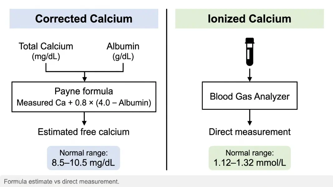
Corrected calcium is calculated by adjusting total serum calcium upward when albumin is below the reference level of 4.0 g/dL. The formula assumes that about 45% of serum calcium circulates bound to albumin. When albumin is low, some of that protein-bound fraction is instead free in solution, making total calcium appear falsely depressed.
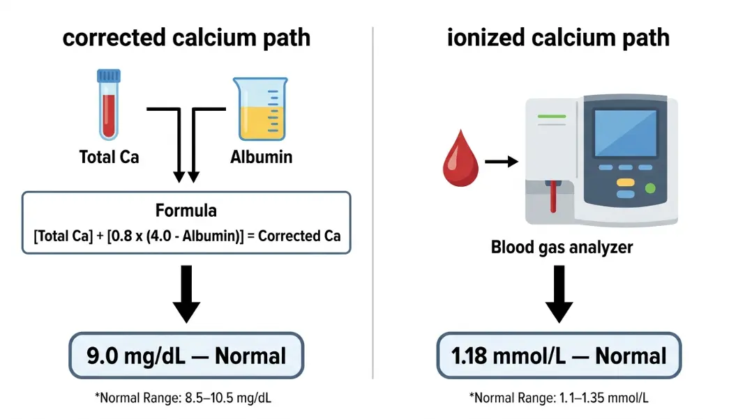
Corrected Calcium (mg/dL) = Measured Calcium + 0.8 × (4.0 - Albumin [g/dL])
Worked example:
- Measured calcium: 7.8 mg/dL
- Albumin: 2.5 g/dL
- Albumin deficit: 4.0 - 2.5 = 1.5 g/dL
- Correction: 0.8 × 1.5 = 1.2 mg/dL
- Corrected calcium: 7.8 + 1.2 = 9.0 mg/dL (within the normal range of 8.5-10.5)
The 0.8 coefficient is a population average for the binding affinity between calcium and albumin. It was derived from a relatively small study of patients who were metabolically stable and had normal acid-base status. That origin matters a great deal once you start applying the formula to patients who are not metabolically stable.
For most outpatient assessments and routine inpatient metabolic panels, the formula performs reliably. The limitations below are situational, not universal.
What Ionized Calcium Actually Measures#
Ionized calcium (Ca2+) is the biologically active fraction. It is not bound to protein. It acts directly on cell membranes, drives cardiac action potentials, controls neurotransmitter release, and triggers muscle contraction. Normal ionized calcium is 1.12 to 1.32 mmol/L (approximately 4.5 to 5.3 mg/dL).
This value is measured directly from a blood gas analyzer or an ion-selective electrode. It is not calculated from total calcium and albumin. The measurement requires a separate specimen handled under anaerobic conditions, analyzed quickly before CO2 shifts alter the result.
The physiological question is always: how much free calcium is available to cells? Total calcium gives an approximation. Corrected calcium gives a better approximation. Measured ionized calcium gives the actual number.
Corrected calcium is used more often in routine practice because it requires no additional test. The albumin and calcium values come from the same standard metabolic panel. Ionized calcium adds cost, time, and specimen handling requirements. For most stable patients, the formula-based estimate is close enough to be clinically useful.
When the Payne Formula Gives a Misleading Result#
Acid-base disturbances. Calcium binding to albumin changes with pH. Alkalosis increases binding, reducing free ionized calcium even when corrected calcium looks normal. Acidosis decreases binding, raising ionized calcium even when total calcium appears low. A patient in metabolic alkalosis can have a genuinely low ionized calcium while the Payne formula reports a normal corrected value. The formula has no pH adjustment. It cannot account for pH-driven shifts in binding.
Critical illness and sepsis. The Payne study was conducted in stable patients. Applying it to critically ill patients with multi-system dysfunction produces errors that several validation studies have documented. Comparisons of corrected calcium to directly measured ionized calcium in ICU populations show misclassification rates of 30 to 60%, depending on severity of illness. This is not a marginal discrepancy. It has direct treatment implications.
Hyperproteinemia. The formula corrects downward for low albumin. It does not correct for elevated protein from paraproteinemias like multiple myeloma. Extra immunoglobulins bind calcium, elevating total measured calcium in ways the Payne formula does not adjust for. A patient with myeloma can appear hypercalcemic on total calcium and normal on corrected calcium when ionized calcium is actually elevated.
Post-parathyroidectomy. After parathyroid surgery, ionized calcium can drop rapidly. Total calcium lags. The corrected calcium formula, applied to a total calcium value drawn before the ionized drop has fully propagated, will overestimate active calcium at precisely the moment accurate measurement matters most.
Neonates and infants. The 0.8 coefficient and 4.0 g/dL reference were derived from adults. Neonatal albumin binding characteristics differ enough that pediatric applications produce less reliable results, particularly in preterm infants where albumin levels, binding affinities, and acid-base status are all atypical.
When to Order Ionized Calcium Instead#
The practical answer: whenever the Payne formula's core assumptions do not hold.
Order ionized calcium directly when any of these apply:
- Acid-base disturbance is present and the clinical picture makes calcium status relevant. Any pH outside 7.35-7.45 with neuromuscular symptoms or cardiac rhythm concerns.
- Critical illness or sepsis. Most critical care guidelines recommend direct ionized calcium measurement as the standard in ICU populations. Corrected calcium should not be the basis for treatment decisions in this setting.
- Symptoms do not match the corrected value. Tetany, perioral paresthesias, Chvostek's sign, or a prolonged QTc interval alongside a normal corrected calcium warrants direct measurement before concluding calcium is not the cause.
- Post-parathyroidectomy monitoring. Direct measurement is standard in the immediate post-operative window when rapid, clinically significant drops are expected.
- Suspected myeloma or paraproteinemia. Total calcium may be elevated from protein binding rather than true hypercalcemia. Direct measurement removes that confounder.
- Neonatal hypocalcemia. Ionized calcium is the preferred measurement in newborns, particularly premature infants.
Outside these situations, corrected calcium using the Payne formula is clinically adequate for outpatient metabolic panels and stable inpatient assessments. The full formula mechanics and clinical interpretation thresholds are covered in the Corrected Calcium Formula guide.
Reading Both Values When They Disagree#
If you have both a corrected calcium from a metabolic panel and an ionized calcium from a blood gas and they do not match, one of three things is happening.
pH shift. Calculate what the ionized calcium should be if corrected calcium is accurate and pH is normal. If measured ionized calcium is lower than expected, alkalosis has increased binding. If higher, acidosis has decreased it. Act on the ionized value in this scenario. It reflects what the cells are actually seeing.
Specimen handling error. Ionized calcium falls if a blood gas specimen sits too long or is exposed to air before analysis. CO2 escapes, pH rises, binding increases, and measured ionized calcium drops. If the ionized value seems inconsistently low and the clinical picture does not support hypocalcemia, repeat the specimen under proper collection conditions.
True discordance from underlying disease. Hypomagnesemia causes functional hypocalcemia at the receptor level despite near-normal measured values. Primary hypoparathyroidism can produce low ionized calcium even when total and corrected values suggest only mild disease. Vitamin D-related conditions sometimes show discordance between total and ionized calcium fractions depending on the binding protein distribution.
In all three scenarios, the ionized calcium is the direct physiological measurement. Corrected calcium is the estimate. When the two disagree, the ionized value is what the biology is actually doing. The discordance itself carries diagnostic information.
For a related electrolyte context, the Anion Gap Calculator identifies the acid-base pattern that is often driving discordance between corrected and ionized values, particularly in patients with an elevated gap pointing toward a metabolic acidosis or in mixed acid-base disorders.
The Formula's Strength Is Its Scope#
None of the limitations above mean the Payne formula is obsolete or unreliable. For stable patients with chronic hypoalbuminemia from malnutrition, liver disease, or nephrotic syndrome, who have no significant acid-base disturbance, the formula remains accurate and clinically useful. It adds real information from a test the patient was already having.
The errors arise from applying a tool validated in one population to a different population. A formula derived in stable, metabolically normal patients will produce less accurate results in critically ill patients with multi-organ dysfunction. That is a scope problem, not a formula flaw.
The working rule: use corrected calcium as your first-pass screen in stable patients. When the clinical context introduces acid-base disturbance, critical illness, paraproteinemia, or any of the conditions above, confirm with direct ionized calcium rather than relying on a correction that was never validated in those settings. The Corrected Calcium Calculator runs the Payne formula for any calcium and albumin values, and the result includes the normal range classification to support that initial screen.
Corrected calcium is an estimate. It adjusts total serum calcium for low albumin using the Payne formula: Corrected Ca = Measured Ca + 0.8 × (4.0 - Albumin). Ionized calcium is a direct measurement of the free, biologically active calcium fraction, performed by a blood gas analyzer or ion-selective electrode. Corrected calcium comes from a routine metabolic panel with no extra specimen. Ionized calcium requires a separate anaerobic specimen handled under specific collection conditions.
Ionized calcium is more reliable than corrected calcium in patients with acid-base disturbances, critical illness or sepsis, after parathyroid surgery, in neonates, and when symptoms suggest hypocalcemia despite a normal corrected value. The Payne formula assumes normal pH and stable albumin binding. Acid-base shifts alter how much calcium binds to albumin, making the calculated correction unreliable without a pH adjustment term that the Payne formula does not include.
Normal ionized calcium is 1.12 to 1.32 mmol/L, equivalent to approximately 4.5 to 5.3 mg/dL. Values below 1.12 mmol/L indicate hypocalcemia. Values above 1.32 mmol/L indicate hypercalcemia. These ranges apply to direct measurement from a blood gas analyzer or ion-selective electrode, not to formula-calculated values. Normal ranges may vary slightly between institutions depending on analyzer calibration.
Yes. Alkalosis increases albumin binding of calcium, pulling ionized calcium down even when total and corrected calcium appear normal. This occurs in respiratory alkalosis from hyperventilation, or metabolic alkalosis from nasogastric suctioning or aggressive diuresis. A patient with tetany, paresthesias, or Chvostek's sign alongside a normal corrected calcium in the setting of alkalosis should have ionized calcium measured directly before calcium status is dismissed as a cause.
The 0.8 coefficient represents the average change in total serum calcium per 1 g/dL change in albumin, derived from regression analysis in the original 1973 Payne study. It reflects albumin-calcium binding affinity under normal physiological conditions. Some clinical protocols use an alternative coefficient of 0.55 when total protein rather than albumin is the measured value. The albumin-based version with 0.8 remains the most widely cited and the version used in most clinical calculators.
Ionized calcium is not calculated from other values. It is measured directly from blood using an ion-selective electrode in a blood gas analyzer or dedicated calcium analyzer. The result represents the concentration of free, unbound calcium ions in the sample at the time of measurement. Some point-of-care blood gas analyzers report ionized calcium automatically alongside pH, pCO2, and pO2. If your lab report shows both total calcium and a separate ionized calcium result, the ionized value is the direct measurement and does not require formula adjustment.
Written by
Hassaan Rasheed
Web Developer & Content Researcher
Hassaan builds calculators and writes source-linked guides across the site's subject areas. Calculator methods and reference data are documented in each guide so readers can verify the underlying sources.
About the author and methods